Detecting Single Proteins By Their Shadows
By Chuck Seegert, Ph.D.

For some time the field of optical sensing has been dominated by fluorescent marker methods. By eliminating these markers, however, a new method from the Max Planck Institute for the Science of Light might significantly advance this field of research.
In many laboratory methods, increasing sensitivity means a smaller sample is required to get reliable results. This is desirable in many areas of medicine — biosensing in particular. For some time, researchers have expanded the limits of optical detection of single molecules by using fluorescent labels to mark proteins of interest. According to a recent study published in Nature Communications, however, eliminating the need to label could increase sensitivity of single-protein detection by many orders of magnitude. This would allow the study of single molecules at the smallest levels of biological matter.
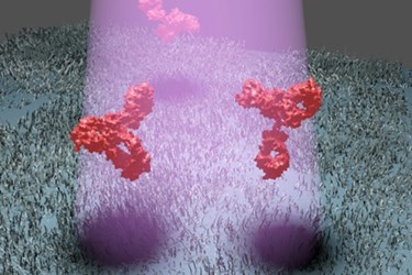
Eliminating fluorescent labels and directly observing proteins has taken a different direction than some expected, according to a recent press release. The new method is referred to as interferomic detection of scattering (iSCAT), and it makes use of a laser that illuminates proteins captured on a slide by biochemical tethers. The laser casts a shadow of the protein, which can then be analyzed.
“Until now it was thought that if you want to detect scattered light from nanoparticles, you have to eliminate all background light,” explained Vahid Sandoghdar, one of the researchers involved with the study, in the press release. “However, in recent years we’ve realized that it is more advantageous to illuminate the sample strongly and visualize the feeble signal of a tiny nanoparticle as a shadow against the intense background light.”
Still, casting a shadow wouldn’t be enough to visualize these single-protein particles without another vital step. First, a baseline image is taken of the iSCAT setup without the sample present. Then, the protein of interest is placed in the field, and another image is taken.
“Since most of the optical noise generated by nanoscopic irregularities of the sample do not change, we can subtract one image from the other and thus eliminate the noise,” said Marek Piliarik, another researcher involved with the study, in the press release.
This makes the shadow from the protein very prominent, making it possible to understand the protein’s structure and functions. This is true even when other proteins are included in the sample, indicating the strong specificity of this method.
The team feels the method has application in many areas of biomedicine.
“iSCAT not only promises more sensitive diagnosis of diseases such as cancers, but will also shed light on many fundamental biochemical processes in nature,” said Sandoghdar in the press release.
Increasing sensitivity and detecting single molecules is an area of research that is undergoing high levels of effort. Recently, a method was developed that allowed for Raman spectroscopy to identify single molecules of material, which is remarkable as this method usually has low sensitivity and requires relatively large samples.
Image Credit: Max Planck Institute for the Science of Light
