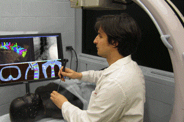
Johns Hopkins Researchers Develop New Image-Based Surgical Guidance System
By Joel Lindsey

A group of researchers at Johns Hopkins University has created a computer algorithm that could give surgeons more precise imaging capabilities to aid in the performance of minimally invasive operations.
“Imaging in the operating room opens new possibilities for patient safety and high-precision surgical guidance,” Jeffrey Siewerdsen, professor of biomedical engineering at the Johns Hopkins University School of Medicine, said in a release issued by the school. “In this work, we devised an imaging method that could overcome traditional barriers in precision and workflow. Rather than adding complicated tracking systems and special markers to the already busy surgical scene, we realized a method in which the imaging system is the tracker and the patient is the marker.”
The process created by Siewerdsen and his team allows C-arm devices, already common in many operating rooms, to create detailed and highly accurate models of the patient’s body by combining data from two-dimensional X-ray images and three-dimensional CT scan images.
This new computerized program could represent a significant improvement over traditional imaging techniques that require a doctor to manually match points on the patient’s body with those depicted in a preoperative CT scan in a process called registration. This process is tedious and prone to human error, both of which may be effectively reduced or eliminated by the new program.
“The registration process can be error-prone, require multiple manual attempts to achieve high accuracy, and tends to degrade over the course of the operation,” said Siewerdsen.
Early studies of the team’s new algorithm report a level of accuracy better than 2 millimeters, compared to the typical accuracy levels of between 2 and 4 millimeters currently achieved by surgical assist devices. Researchers believe this increased accuracy may translate into improved navigational capabilities for surgeons while performing operations.
“We are already seeing how intraoperative imaging can be used to enhance workflow and improve patient safety,” said Ziya Gokaslan, a professor of neurosurgery at the Johns Hopkins University School of Medicine who is also involved with the project. “Extending those methods to the task of surgical navigation is very promising, and it could open the availability of high-precision guidance to a broader spectrum of surgeries than previously available.”
Details of the project have been released in an article published recently by the journal Physics in Medicine and Biology.
Researchers involved with the project are currently preparing the computer program for use in clinical studies, according to Johns Hopkins. At this point, Siewerdsen foresees the technique being used primarily in minimally invasive surgeries such as spinal and intracranial neurosurgery.
Image credit: Johns Hopkins Medicine
