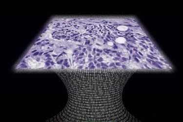
Lens-Free Microscope Could Lead To Cheaper, Portable Imaging Tech
By Chuck Seegert, Ph.D.

Research from the University of California at Los Angeles (UCLA) has led to a new, lens-free microscope that can detect cellular abnormalities like cancer. The portable system has a similar accuracy to the larger, more expensive equipment often found in pathology labs, so it may enable diagnostics in more remote areas.
The pathological examination of tissues is essential for biopsies in cancer diagnostics. Traditionally, light-based microscopes have been used to examine tissue specimens and blood samples, and this has been the case for several decades. While effective, these light-based microscopes are limited because of their relatively high-cost and lack of portability. Additionally, the physics of light limits the field-of-view, which requires a researcher to scan a slide by moving it along two axes to study the entire specimen. As the slide is translated like this, it may require 3D focus adjustment along the way.
Many of these limitations may now be overcome based on work from researchers at UCLA, according to a recent article from the UCLA Newsroom. The new, lens-free design is the latest example of a computational diagnostic device that has come from the lab of Aydogan Ozcan, the Chancellor’s Professor of Electrical Engineering and Bioengineering at the UCLA Henry Samueli School of Engineering and Applied Science.
“This is a milestone in the work we’ve been doing,” Ozcan said in the press release. “This is the first time tissue samples have been imaged in 3D using a lens-free on-chip microscope.”
In the newly developed device, light-emitting diodes (LEDs) or lasers are used to illuminate the specimen which is inserted into the instrument. Using an on-chip sensor array similar to those used in cellphone cameras, the patterns of shadows cast by the specimen are recorded. The system generates patterns in a holographic form and provides the user with a virtual depth-of-field. The images are adjusted automatically to enhance contrast in the specimen, which makes abnormalities easier to detect.
When a pathologist used images from the new device to analyze breast cancer cells, Pap smear specimens, and sickle cell blood samples, results were ~99 percent accurate, according to a recent study published by the team in Science Translational Medicine. The system digitally corrects for possible tilts or differences in the planes between the specimen and the sensor, and it doesn’t need to be manually refocused. Additionally, the field-of-view is much greater than traditional microscopes and spans an area of about 20.5 square millimeters. The image can also be digitally refocused at any depth in the sample.
The need for high-quality pathological analysis is often found in areas where laboratory infrastructure is poor, such as third-world environments. Other researchers are also working to increase portable capabilities for alternative laboratory needs. For example, a portable mass spectrometer was recently developed to perform analysis that is usually confined to large institutional labs.
Image Credit: UCLA Newsroom
