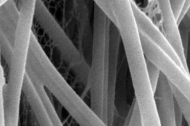
Anti-Inflammatory Nanofibers Help Advance Tissue Regeneration
By Chuck Seegert, Ph.D.

The implantation of scaffold materials is frequently associated with inflammatory reactions in the area surrounding them. To mitigate the negative effects of inflammation and enhance the performance of these implants, researchers at Ann & Robert H. Lurie Children's Hospital of Chicago have developed a nanofiber anti-inflammatory approach.
Inflammation is a normal response involved in the healing process. It assists in the regeneration of tissue and helps banish infection, though it may be somewhat painful. The same mechanism, however, can have a negative impact when treatment includes an implant, as the body will also marshal a response against the implant. In some situations, this has less of an impact on implants made of materials like titanium or other metals, but in other cases, it can be detrimental to the implanted device.
The use of biologically based materials can induce inflammation, and when this happens, tissue fibrosis, or stiffening, can occur. Stiffening of these biologics can interfere with the properties that make them attractive in the first place, forcing them to behave differently than the original tissue. To combat these physiological reactions, a research team led by Arun Sharma, Ph.D., has formulated anti-inflammatory nanofibers that have been incorporated into a scaffold.
Sharma is the director of pediatric urological regenerative medicine at Ann & Robert H. Lurie Children's Hospital of Chicago, the director of surgical research at Stanley Manne Children’s Research Institute, an assistant professor in the departments of urology and biomedical engineering at Northwestern University’s Feinberg School of Medicine, and a member of the developmental biology program of the research institute.
In a research study published in Biomaterials, Dr. Sharma and his group looked at an existing bladder augmentation model composed of decellularized small intestine submucosa (dSIS). Historically this material has been highly inflammatory, but when treated with self-assembling, anti-inflammatory peptide amphiphiles (AIF-PAs), the results were much different.
Treatment with AIF-PAs demonstrated several regenerative advantages including an elevated angiogenic response, indicating that the material was being vascularized differently than before. Additionally, little collage accumulation was seen, and the presence of inflammatory M2 macrophages were reduced compared to the control group. Cytokines, or cell-signaling molecules associated with inflammation were decreased, while anti-inflammatory cytokines had increased.
“Our findings are very relevant not just for bladder regeneration but for other types of tissue regeneration where foreign materials are utilized for structural support. I also envision the potential utility of these nanomolecules for the treatment of a wide range of dysfunctional inflammatory based conditions,” said Sharma in a recent press release.
The use of nanofibers in tissue engineering is an area of research that is receiving increasing levels of focus. Recently, another nanofiber technology was discussed for tissue engineering and drug delivery applications.
