3D Printing Enables Tailor-Fit, Sensor-Loaded Heart Wrap
By Joel Lindsey

A team of biomedical engineers has used 3D printing to create a highly customizable implantable device, embedded with a series of sensors, that could significantly alter the way doctors diagnose, monitor, and treat heart disorders.
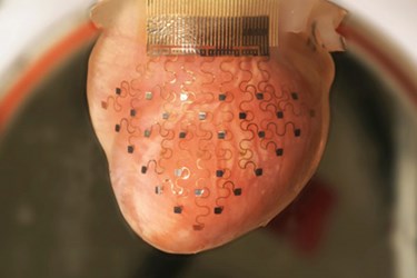
Using data computationally extracted from MRI and CT scans, the researchers can print an exact plastic replica of a patient’s heart. They can then use that model to mold a flexible membrane that will adhere directly to the patient’s actual heart. An arrangement of sensors capable of measuring temperature, mechanical strain, and pH, as well as delivering pulses of electricity is then embedded into the soft membrane.
By wrapping the membrane around the outer wall of a patient’s heart, it is hoped that surgeons and doctors can use the data gathered by the sensors to monitor heart treatment, manage arrhythmia, and more accurately predict heart attacks, a press release published by Washington University in St. Louis reported.
“Each heart is a different shape, and current devices are one-size-fits-all and don’t at all conform to the geometry of a patient’s heart,” said Igor Efimov, Lucy & Stanley Lopata Distinguished Professor of Biomedical Engineering at Washington University and co-leader of the research team developing the new technique.
Details regarding early developments of this technique have recently been published online in the journal Nature Communications.
At this point, researchers have said that the device could be used to treat diseases or disorders affecting the ventricles in the lower chambers of the heart. It could also be used to assist in the treatment of atrial fibrillation.
“Currently, medical devices to treat heart rhythm diseases are essentially based on two electrodes inserted through the veins and deployed inside the chamber,” Efimov said. “Contact with the tissue is only at one or two points, and it is at a very low resolution. What we want to create is an approach that will allow you to have numerous points of contact and to correct the problem with high-definition diagnostics and high-definition therapy.”
Scientists hope to eventually see this technique extended to fields beyond strictly cardiac treatment.
“Because this is implantable, it will allow physicians to monitor vital functions in different organs and intervene when necessary to provide therapy,” Efimov said. “In the case of heart rhythm disorders, it could be used to stimulate cardiac muscle or the brain, or in renal disorders, it would monitor ionic concentrations of calcium, potassium, and sodium.”
3D printing is increasingly being explored as a possible tool in cardiac medicine. Earlier this month, surgeons in Louisville, Kentucky used 3D printing to create a detailed model of an infant’s heart. The model then allowed them to make more accurate diagnoses and to plan out and practice beforehand how to perform the complex surgeries required for the young boy’s treatment.
Image Credit: Igor Efimov
