New MRI Approach Could Detect Prostate Cancer More Accurately
By Chuck Seegert, Ph.D.

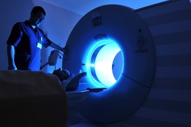
To overcome challenges with existing magnetic resonance imaging (MRI) methods, researchers at the University of California have developed a new, more accurate approach to imaging prostate cancer. By correcting magnetic field distortions, the new method may significantly change how prostate cancer is diagnosed and treated.
Detecting prostate cancer is currently done through contrast enhanced MRI. A contrast agent is injected into the patient’s blood stream, and it highlights any abnormalities that may be present as it passes through tissue. In tumors, rapidly growing cells often need a greater amount of blood to support them compared to the healthy tissue surrounding the cancer. This may illustrate the location of the tumor, but it is not a guaranteed process. Many tumors are not distinct enough to be noticed in the images.
Another method emerging in the diagnosis of prostate cancer is called diffusion MRI, which has historically been a standard-of-care in brain imaging. This method examines differences in water between denser tumors, which have less water content than normal tissues. The problem with this method, however, is that magnetic field distortions can show tumor locations in the images that may be up to 1.2 centimeters from their actual locations, according to a recent article from the UC San Diego Newsroom. This can be significant when physicians are trying to evaluate if the tumor is affecting dense bundles of nerves in the area.
To improve this process, a joint team from the University of California, San Diego (UCSD) and University of California, Los Angeles (UCLA) has developed a new method called restriction spectrum imaging-MRI (RSI-MRI), according to a recent study published by the team in the journal Prostate Cancer and Prostatic Diseases. The new method corrects for the distortions in the magnetic field, and also focuses on water diffusion, thus increasing accuracy. In a sample of 27 patients, the RSI-MRI method was able detect extraprostatic extension of the cancer (growth outside the organ) 89 percent of the time, while traditional MRI only detected it 22 percent of the time.
In addition to detecting cancer more accurately than standard MRI approaches, the new technique may be able to detect the stage of the disease, according to the article. High-grade tumors appear to have a more restricted water volume than lower grade tumors when studied by SRI-MRI. If detection of the tumor grade could be accomplished non-invasively, some patients could be monitored without resections or biopsies.
Non-invasive detection of cancers and other diseases holds much attraction in the diagnostic space. One example of a new technique to detect prostate cancer uses a biomarker secreted in the urine, which is analyzed using an electronic nose.
