New Single-Chip Device May Provide Internal 3D Imaging Of Cardiovascular System
By Joel Lindsey

Researchers at the George W. Woodruff School of Mechanical Engineering at Georgia Institute of Technology have created a tiny single-chip device that may give doctors and surgeons a real-time, three-dimensional internal view of patients’ blood vessels and hearts.
According to the researchers, the device could significantly improve cardiologists’ ability to treat blocked arteries and other related health problems.
“Our device will allow doctors to see the whole volume that is in front of them within a blood vessel,” professor and project researcher F. Levent Degertekin said in an article published by Georgia Tech. “This will give cardiologists the equivalent of a flashlight so they can see blockages ahead of them in occluded arteries. It has the potential for reducing the amount of surgery that must be done to clear these vessels.”
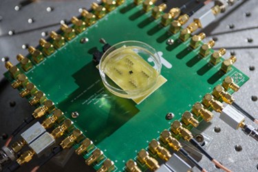
The device measures 1.5 millimeters in diameter and includes a 430-micron hole in the center to accommodate a guidewire. The device is designed to be inserted directly into blood vessels with a catheter, where it then uses a combination of capacitative micromachined ultrasonic transducer (CMUT) arrays and front-end complementary metal-oxide semiconductor (CMOS) technology to generate 3D intravascular ultrasound (IVUS) and intracardiac echography (ICE) images.
The device also includes power-saving circuitry that shuts sensors down when they’re not in use. This allows the chip to operate on 20 milliwatts of power, thereby limiting the amount of heat generated inside the body.
“If you’re a doctor, you want to see what is going on inside the arteries and inside the heart, but most of the devices being used for this today provide only cross-sectional images,” Degertekin said. “If you have an artery that is totally blocked, for example, you need a system that tells you what’s in front of you. You need to see the front, back and sidewalls altogether. That kind of information is basically not available at this time.”
Researchers have so far built and tested a prototype capable of generating visual data at a rate of 60 frames per second. Their next focus will be testing the device in animals. Ultimately, they expect to license the technology to an already-established medical diagnostic firm to perform whatever clinical trials are necessary for FDA approval.
According to the Georgia Tech article, Degertekin and his team are already thinking about additional applications for the device.
“Degertekin hopes to develop a version of the device that could guide interventions in the heart under magnetic resonance imaging,” the article reported. “Other plans include further reducing the size of the device to place it on a 400-micron diameter guide wire.”
Details from this research have been published in this month’s edition of IEEE Transaction on Ultrasonics, Ferroelectronics and Frequency Control.
Image Credit: Rob Felt, Georgia Tech
