Researchers Fuse Imaging Methods For Greater Surgical Precision

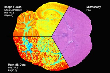
Researchers have created a predictive imaging modality by combining imaging mass spectrometry (IMS) and microscopy. Because the two technologies have their own unique advantages, researchers say the new technology can provide more specific information at a higher resolution, allowing clinicians to make more informed diagnostic and treatment decisions.
According to Richard Caprioli, senior author of a paper published in Nature Methods, microscopy provides excellent spatial information and high-resolution imaging of tissue, while IMS-generated molecular maps are rich in chemical specificity. By combining the two, Caprioli says clinicians are able to achieve the best of both worlds.
A team at Vanderbilt University used a mathematical approach called regression analysis to map each pixel of the mass spectrometry image onto the corresponding location on the microscopy image. Therefore, scientists were able to “model variables in one technology, using variables from the other technology.” This process creates what researchers call a “predicted image.”
In the study, the authors demonstrated the potential of the new technology by “sharpening IMS images, which uses microscopy measurements to predict ion distributions at a spatial resolution that exceeds that of measured ion images by ten times or more, predicting ion distributions in tissue areas that were not measured by IMS, and enriching of biological signals and attenuation of instrumental artifacts, revealing insights not easily extracted from either microscopy or IMS individually.”
In a way, said Caprioli in a Vanderbilt research news story, “we’re predicting what the data should look like. It is an important step in the process of making mass spectrometry data accessible and truly useful for clinicians.”
Caprioli projects that the technology could prove useful for surgeons attempting to resect cancerous tumors by giving them a more precise “margin” between normal and cancerous cells to follow.
Typically, Caprioli explains, cancerous cells are located using microscopy, which doesn’t give a complete picture of their protein content. Without the additional information provided by IMS technology, many developing cancerous cells may be overlooked. Caprioli thinks this is one of the reasons why many cancers recur.
“The application of image fusion approaches to the analysis of tissue sections by microscopy and mass spectrometry is a significant innovation that should change the way that these techniques are used together,” said Douglas Sheeley, senior scientific officer in the National Institute of General Medical Sciences (NIGMS).
The research was funded by National Institute of Health (NIH) grants.
Image credit: Vanderbilt University
