Siemens, U. of Twente Biopsy Robot Promises Greater Precision, Less Cost

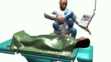
A collaboration between European academic and industry scientists has developed a surgical robot that combines magnetic resonance imaging (MRI) and ultrasound technology to improve biopsy precision and reduce the necessity for expensive and time-consuming medical imaging. The research team is currently testing and refining the technology, which they say could be used to diagnose breast cancer and muscle diseases.
The MRI and Ultrasound Robotic Assisted Biopsy (MURAB) project is centered at the University of Twente (UT), where a research team led by Foad Sojoodi Farimani and Stefano Stramigioli is collaborating with scientists from Siemens and KUKA Robotics, as well as universities in Verona and Vienna.
MURAB is aimed at guiding surgical biopsies so doctors can be sure they’re analyzing the best possible sample. “If mammography shows a suspicious image, a tissue sample is taken and sent to the laboratory for analysis. But it is difficult to determine exactly where the biopsy must take place,” explained Farimani in a press release. “Far too often cancers are overlooked. This is the challenge we want to address.”
According to Farimani, MRI technology provides the sharpest and most accurate images of suspicious tissue with the least risk to the patient; however, these tests take a lot of time — between 45 and 60 minutes, if not longer — and are prohibitively expensive for the most well-funded and developed healthcare systems. The MURAB, by comparison, completes the MRI scan portion of the biopsy in about 15 to 20 minutes.
The system works by first taking an MRI image and then overlaying it with an image taken by ultrasound and pressure sensors during the biopsy — using the sharper image to locate suspicious tissue within the less distinct ultrasound. Working from the combined images, surgeons will be able to remove more precise samples, said Farimani.
The team anticipates that MURAB technology will be useful for any procedure where a precise tissue sample is needed. Besides cancers, the system will also be able to diagnose certain muscle diseases. Currently, the system has a 10 to 20 percent failure rate, meaning it misses cancers 1 out of 5 times — a success ratio the team is working to improve.
The MURAB project recently received $4.6 million from Horizon 2020, a European commission launched in 2014 that will fund $87 billion worth of research through 2020. A quarter of the $4.6 million will be put toward management and commercialization of the project.
“The robotics in this project aren’t the hard part,” Farimani told Oost NV. “But making sure the resulting technology has a good fit to the market — that’s easier said than done.”
UT recently hosted a symposium on the European robotics industry, which International Innovation reports is experiencing an 8-percent-per-year growth rate. Stramigioli remarked that European success with innovative robotic technology was due to strong international collaboration.
“Europe has incredible potentials in robotics and can become the leader in different relevant applications,” said Stramigioli. “The way we work together with European teams in Europe is pretty unique.”
WinterGreen market researchers estimate that the market for surgical robotics will reach $20 billion by 2021.
