UVA Scientists Combine MRI, Gamma Rays For HQ Medical Diagnostics

A new medical imaging modality developed by scientists at the University of Virginia (UVA) combines magnetic resonance imaging (MRI) with gamma-ray imaging using xenon isotopes. UVA researchers call polarized nuclear imaging (PNI) an “exciting new technology” that combines the spatial and spectral advantages of MRI with radioactive tracers to produce diagnostic information previously unavailable.
PNI works using radio-frequency electromagnetic radiation and magnetic-field gradients — similar to MRI — but instead of a radio wave detector, the system instead detects gamma rays given off by radioactive tracers. In a biological subject, researchers explained that the isotopes could either be inhaled or injected near the examination site.
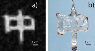
Wilson Miller and Gordon Cates, UVA physicists and co-directors of the research, demonstrated their PNI system using a small glass cell filled with a trace amount of xenon that had been polarized using laser techniques. Xe-131m is a by-product of Iodine 131, which is used to treat thyroid problems, and the trace amounts that would be used for medical imaging pose little to no risk to patients. Their results — published in Nature — produced images with isotopes using a fraction of the amount of material found in the water required by conventional MRI, which would require 10 billion more atoms to produce an image of comparable quality.
“Unlike MRI, which detects faint radio waves, we detect gamma rays that are emitted from the xenon isotope,” Cates said in a press release. “Since it is possible to detect a gamma ray from even a single atom, we gain an enormous increase in imaging sensitivity and dramatically reduce the amount of material needed for performing magnetic-resonance techniques.”
Authors in the study noted that the high sensitivity offered by the new modality greatly expands potential applications for MRI, and may lead to a new approach in the development of radioactive tracers. This technique could potentially yield diagnostic information that is impossible using other methods, and researchers speculated that it may be especially effective in imaging lung tissue.
“This method makes possible a truly new, absolutely different class of medical diagnostics,” said Miller. “There was once a first x-ray image, and a first CT-scan image, and a first MRI image. We now have produced the first image of a new technology, PNI, which someday may be as much in use as those others.”
Vanderbilt scientists introduced a predictive imaging modality last year that combined imaging mass spectrometry (IMS) and microscopy for greater surgical precision. Microscopy offers spatial information and IMS offers chemical breakdowns of the tissue, which is useful when distinguishing between healthy and cancerous tissue, said researchers.
In August, the FDA approved Siemens Healthineers’ Somatom Drive computed tomography (CT) system, which allows for higher resolution images with lower doses of contrast and radiation. The system allows for multiple kV settings, allowing clinicians to tailor imaging to the specific indication and patient.
