Fiber Optics May Provide Minimally Invasive Alternative To Traditional Muscle Biopsy

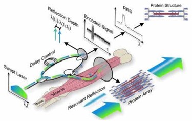
New technology may provide a minimally invasive alternative to traditional muscle biopsies, which are commonly used to diagnose patients showing symptoms of muscular disorders, diseases, or infections. The technique uses reflection spectroscopy (RSS) to image nanoscale muscle movements impossible to study with traditional methods, and could lead to a better understanding of degenerative diseases, such as Muscular Dystrophy, Lou Gehrig’s Disease, stroke, and Parkinson’s.
Muscle biopsies , which are the current gold standard in muscle diagnostics, are taken using a biopsy needle, or through a small incision at the location of symptoms. While a study of individual muscle cells can provide some diagnostic information, the procedure is damaging to the living tissue, painful and risky to the patient, and does not yield much information about muscle movement. Newer and less invasive advances in biopsy, such as laser diffraction and microendoscopy, are likewise limited in scale and ability to capture small movements.
According to scientists from the Rehabilitation Institute of Chicago (RIC), the best way to assess muscle health is to measure sarcomeres, which are responsible for generating muscle force. Sarcomeres are the basic unit of muscle tissue, and there are approximately 100,000 sarcomeres in one muscle fiber.
In a study published in Biophysical Journal, RIC scientists demonstrated that RSS using a fiber optic probe 250μ-wide is more precise and less invasive that current methods in assessing nanoscale changes in sarcomere length. Researchers were able to capture length changes in thousands of sarcomeres on a sub-millisecond timescale during passive stretch-and-twitch contraction of a rabbit’s leg muscle in vivo.
Richard Lieber, physiologist and senior author of the study, explained in a press release that “This approach enables measurement of previously unobtainable muscle properties by combining advances in telecommunications technology with a deep understanding of muscle structure, biomechanics, and pathology... To our knowledge, this method achieves sample sizes, resolutions, and compatibility with human movement that no other current or proposed technique can match.”
Lieber and his team hope to continue refining the technology to make it more accessible to researchers worldwide, and to continue using the technology to generate new research on muscle movements, pathologies, and the effectiveness of different therapies on muscle health.
