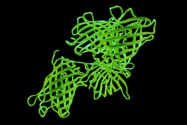
Fluorescent Protein Optical Imaging Considerations

Fluorescent proteins (FPs) have significantly advanced scientific fluorescence applications, particularly in microscopy, which typically uses light from ultraviolet to infrared wavelengths. While most studies focus on the visible spectrum, there is growing interest in far-red FPs, as they cause less cellular damage. Key factors in utilizing FPs include image acquisition speed, which necessitates high sensitivity detectors and bright light sources for live cell imaging. Optimal filter sets with steep spectral edges and high transmission rates are essential to minimize phototoxicity and photobleaching during excitation.
Maintaining live cell conditions—37°C, controlled CO2 levels, and humidity—requires reliable optical components. Hard-coated filters are preferred due to their durability under these conditions. For applications like optical highlighters, multiple excitation bands are needed, necessitating the use of dichroic beamsplitters and filters that effectively manage overlapping optical paths. Careful intensity optimization is crucial to prevent cellular damage from UV light.
In multicolor imaging, such as with Brainbow mice, emission filters must have high transmission rates and narrow passbands to differentiate between various fluorescent signals. Color-corrected objectives are also critical, as they ensure accurate signal registration across different hues. Overall, the careful selection of filters, detectors, and other optical components is vital for maximizing the effectiveness of fluorescent proteins in biological research and imaging.
Get unlimited access to:
Enter your credentials below to log in. Not yet a member of Med Device Online? Subscribe today.
