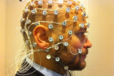
Measuring "Brain Tsunamis" With EEG
By Chuck Seegert, Ph.D.

Spreading depolarizations, or “brain tsunamis,” may now be measurable without surgically opening the skull. Historically, leads have been placed into a patient’s brain tissue, but thanks to advances from the University of Cincinnati (UC), this may no longer be required.
Spreading depolarizations (SD) are waves of self-propagating activity in brain tissues that travel through adjacent neurons. This phenomenon is seen in traumatic brain injury (TBI), as well as migraines and strokes. Essentially, there is a rapid and massive translocation of the ions between the neurons and the extracellular space, which can cause edema and cell death in the brain’s grey matter.
To understand the development of SD, neurosurgeons have to insert leads into the brain tissue directly, a procedure that involves opening the skull and exposing the brain, according a recent UC press release. Spreading depolarizations have been associated with worse outcomes for patients who have had the misfortune of experiencing TBI. Currently only 10 to 15 percent of patients who have had a TBI are subjected to brain surgery and intra-cranial monitoring, according to the press release. This leaves the remaining 85 to 90 percent without options when it comes to studying SD.
Having this information would help guide treatment choices for this large group of patients, if it were available.
"This landmark observation broadens the spectrum of patients and conditions that can be monitored for the occurrence of spreading depolarizations,” said co-investigator Norberto Andaluz, M.D., associate professor of neurosurgery and director of the UC Neurotrauma Center at the UC Neuroscience Institute, in the press release. “Until now, spreading depolarizations could only be detected through the insertion of surgical leads into the patient's brain. Thanks to this discovery, we will acquire a better understanding of the role that depolarizations play in virtually every neurological condition, thus opening new opportunities for therapeutic interventions.”
A study performed by the team was recently published in the Annals of Neurology, and included 18 patients who were followed for 65.9 days using a conventional electroencephalograph (EEG). What’s different from the past, however, was how the team viewed the data, which was compressed to show many hours of EEG recordings at once. Traditionally, neurologists have only looked at the data at 10 second intervals. This change leads to a much different understanding of brain activity.
“The pattern jumps out at you,” said professor Jed Hartings who led the research team, in the press release. “Suddenly, what you see developing over a course of 45 minutes is a depression of the amplitude of the brainwaves. It takes maybe 15 minutes for that to develop to its minimum, and then it will slowly recover again over another 15 to 20 minutes. So you can only see the depolarization when you are looking at long epochs of data. This is something that people have probably been recording for 50 years, but weren’t looking at the data in the right way to be able to recognize it.”
Treatment of TBI is a major focus in today’s healthcare environment, particularly for military occupations, where TBI can be more frequent. According to an article on Med Device Online, the Department of Defense recently kicked off an initiative to develop a rapid point-of-care test for TBI with Abbott Laboratories.
Image Credit: "EEG recording" by Petter Kallioinen. Licensed under Creative Commons Attribution-Share Alike 3.0 via Wikimedia Commons: http://creativecommons.org/licenses/by-sa/3.0/
