Medtronic, WUSM, NIH Collaborate To Better Map And Excise Brain Tumors
By Jof Enriquez,
Follow me on Twitter @jofenriq

Medtronic and researchers at Washington University School of Medicine in St. Louis are using $3.6 million in funding from the National Cancer Institute of the National Institutes of Health (NIH) to build advanced surgical planning software based on customized 3D magnetic resonance imaging (MRI) brain scans.
Typically, patients undergo MRI imaging before neurosurgery to map the brain's anatomical features in and around the tumor site to avoid removing portions of the brain responsible for key functions, such as language or vision. However, it's not quite as straightforward, because individuals vary in where such functions are located. Thus, to map these functions, doctors instruct patients who are awake during surgery to perform simple tasks – such as repeating a word during electrocortical stimulation to identify specific language regions.
“Every neurosurgeon uses a navigation system, but we want it to be even better,” said Eric Leuthardt, MD, a professor of neurosurgery and of mechanical engineering and applied science, and the co-principal investigator on the NIH grant, in a news release. “We want to give them the ability not only to navigate the brain anatomy, but to know the implications of making incisions into each of those components of anatomy.”
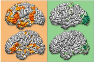
The researchers plan to create a software program based on resting-state functional MRI (rsfMRI), a non-invasive imaging technique that can also map brain function, rather than just brain structure alone. This type of imaging can reveal patterns of brain activity even in periods of sleep – resting states that are thought to reveal different regions in the brain that often act together. Therefore, it does not rely on the patient to follow explicit tasks during brain surgery to map the brain.
Leuthardt and co-principal investigator Joshua Shimony, MD, an associate professor of radiology, worked previously with Medtronic to integrate resting-state data with the company's StealthStation navigational system to make individualized structural and functional maps.
But the implementation was not fully completed and requires a full-time human specialist to produce 3D maps. Now, MD/PhD student Carl Hacker, PhD, has developed an algorithm to crunch and analyze functional data automatically.
“We’re creating a software package that is going to combine our expertise in interpreting resting state data with the navigational technology at Medtronic, so that anyone who wants to use resting state MRI to map functional areas, or to make a brain map for use in surgical planning, can have that readily available,” Leuthardt said. “We think it will be a win-win situation.”
