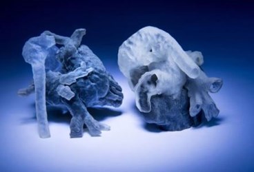
MIT Building A More Accurate 3D Printed Heart For Safer Surgeries

Cardiovascular surgeons have been using 3D-printed heart models crafted from their patients’ MRI images to help plan their surgeries, to anticipate problems, and to speed up their procedures. Models currently take upwards of 10 hours to generate and they are not as accurate as they could be, but MIT scientists believe they can significantly reduce production time, while improving accuracy, by combining computer algorithms with human expertise.
Traditionally, surgeons had to wait until after the first cut to begin planning their strategies but, by then, the patient was under anesthetic, and every minute was critical. Recent advances in 3D printing allow surgeons to handle and examine a virtual copy of their patient’s heart before going into the operating room, a process that experts believe saves valuable minutes.
Last year, a team of cardiovascular specialists at Kosair Children’s Hospital in Louisville, Ky. used such a model to plan surgical intervention on a 14-month-old child with multiple heart defects. Earlier this year, a team at the Children’s Hospital of Los Angeles (CHLA) used the same approach to treat a child with ventricular septal defect, and a team at the Children’s Hospital of Michigan used to a model to practice a risky operation on a teenager’s damaged aorta.
Richard Kim, a cardiac surgeon at CHLA, commented that the model helped to streamline his surgery. “Instead of opening the chest and making a decision about how to proceed, I could immediately begin fixing the problem,” said Kim.
Surgeons who have used the models agree that they save time in the operating room, and less time equals better, safer surgeries. Massachusetts Institute of Technology (MIT) scientist Polina Golland wondered if that time might be even further reduced by developing faster methods for producing the models.
According to MIT News, previous methods required technicians to manually analyze and indicate boundaries on 200 or more cross sections generated by MRI data, a process that can take hours to complete. Efforts to produce a fully automated process relied on generic heart models to fill in gaps of missing information, which compromised the accuracy of the model.
To solve these problems, Golland’s team developed a system in which experts and computers worked together. Their algorithm required experts to identify a fraction of the necessary 200 boundaries and let a computer take over to do the rest. Researchers found that after they segmented only 14 boundaries, the computer was 90 percent in agreement with experts on the remaining 186.
Andrew Powell, a cardiologist at Boston Children’s Hospital (BCH) who’s leading clinical studies for Golland’s model, told the Boston Herald that the team plans to reduce the model’s production time to 30 minutes or less.
Though surgeons are enthusiastic, there still is not a lot of clinical data to support whether 3D heart models improve surgical outcomes. So, this fall, Golland’s team plans to launch a study with seven cardiac surgeons at BCH who will evaluate the device for clinical usefulness.
“Our collaborators are convinced that this will make a difference,“ said Golland to MIT News. “The phrase I heard is that ‘surgeons see with their hands,’ and that perception is in the touch.”
