MIT's Electrode Array Can Monitor Dopamine Levels Long-Term

Scientists from the Massachusetts Institute of Technology (MIT) have developed an electrode array that allows for long-term monitoring of dopamine in the brain, which could shed light on diseases like Parkinson’s and depression, and lead to more effective treatments. While current electrode arrays suitable for dopamine monitoring can last for just a day, MIT’s technology can continue transmitting signals for two months.
Inside the nervous system, Dopamine is a neurotransmitter that plays a role in motor control, reward-based learning, and the release of certain hormones. Many addictive drugs are associated with elevated levels of dopamine, and the dysregulation of the chemical has been linked to several brain disorders. A clear understanding of dopamine and its function within the nervous system has been limited by the technology available to monitor its levels and changes over time.
According to MIT scientists, led by bioengineer Michael Cima, current electrode arrays capable of monitoring dopamine release are approximately 100 microns in diameter, are difficult to target in areas of the brain responsible for dopamine production, and commonly are rendered useless by scar tissue and biofilms after about a day. The scientists’ goal, recently described to MIT News, was to overcome these obstacles that limit the amount of useful information researchers can collect about the function of dopamine and its association with memory, learning, and emotion.
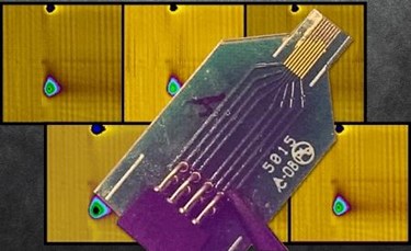
The electrode array developed by MIT is approximately 10 microns in diameter — similar in size to the neurons themselves — and is arranged in groups of eight and coated with polyethylene glycol (PEG) polymer, which protects the delicate electrodes during insertion, then melts away once the device is in place. Oscillating voltage transmitted through the electrodes causes an electrochemical change in the dopamine, which then sends a signal back to be recorded.
In a study published in Lab on a Chip, researchers were able to record dopamine from 16 sites on a rat’s striatum, the portion of the brain responsible for habit formation and reward-based learning. Researchers demonstrated “high density mapping” of dopamine using minimally invasive probes that preserved the viability of surrounding neurons for up to 2 months.
“Nobody has really measured neurotransmitter behavior at this spatial scale and timescale,” said Cima. “Having a tool like this will allow us to explore potentially any neurotransmitter-related disease.”
Helen Schwerdt, a post-doctorate MIT fellow and lead author of the study, explained that deep brain stimulation — which currently is being used to treat the motor symptoms of Parkinson’s disease—is believed to resupply the brain with dopamine but no one really has a clear understanding of how that occurs. According to Schwerdt, this new technology could provide researchers with a clearer understanding of dopamine, and potentially lead to better treatments for neurotransmitter-related conditions.
Since 2013, the National Health Institute, as well as other government agencies, has been funding neuro research that accelerates the development and application of new neuroethologies through the BRAIN Initiative. Technology recently introduced by Harvard Scientists may one day simulate vision in the blind with a “hair-like” implant made from microcoils.
