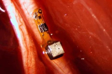
Neural Dust Uses Ultrasound To Monitor Nerves In Real-Time

Scientists at the University of California, Berkeley (UCB) have shrunk biosensors to the size of a single grain of sand and, applying ultrasound to those sensors, can monitor nerves, muscles, or organs in real time. Successful in vivo studies using this “neural dust” in animals are the latest advance in the Defense Advanced Research Projects Agency ‘s (DARPA) Electrical Prescriptions (ElectRx) program, aimed at developing nanoscale devices for the minimally invasive management of chronic illness.
The UCB system provides electromyography (EMG) and electronystagmogram (ENG) recordings from a completely passive device that requires no batteries or wires to connect the device to surrounding tissues, the researchers told Berkeley News. Each sensor is approximately one millimeter across and contains a piezoelectric crystal that converts ultrasound vibrations from outside the body into electricity to power the device. Nerve activity picked up by recording electrodes then changes the current and the vibration of the crystal, which then reflects the data back to the ultrasound receiving device.
The key to the device’s high-quality recordings is the use of ultrasound instead of radio frequency (RF), commonly used in similar devices. RF telemetry requires a great deal of power and suffers significant signal loss through biological tissue, while ultrasonic communication does not. Because they require less power, ultrasonic biosensors can be made much smaller and placed deeper inside the body, said the scientists.
“Neural dust represents a radical departure from the traditional approach of using radio waves for wireless communication with implanted devices,” said David Weber, project manager of ElectRx at DARPA, in a release. “By using ultrasound to communicate with the neural dust, the sensors can be made smaller and placed deeper inside the body, by needle injection or other non-surgical approaches.”
In a study published in Neuron, UBC scientists — led by Michel Maharbiz and Jose Carmena — tested their tiny sensors in the nerve fibers of rats. Using several sensors at once, the scientists were able to achieve high-fidelity recordings from many different sites within a nerve bundle, and they claim their results “highlight the potential” for ultrasound technology in the future of bioelectronics-based therapies.
“I think the long-term prospects for neural dust are not only within nerves and the brain, but much broader,” said Mahrabiz. “Having access to in-body telemetry has never been possible because there has been no way to put something super tiny super deep. But now I can take a speck of nothing and park it next to a nerve or organ, your GI tract or a muscle and read out the data.”
To date, DARPA has funneled $78.9 million dollars into the ElectRX program and has provided grants to seven research projects that submitted proposals last year. According to Weber, the project is seeking to reframe the way we approach healthcare by viewing the human body as a system of electrical circuits.
DARPA recently provided $7.5 million in funding for Profusa’s implantable biosensor technology, capable of transmitting metabolic chemistry data for up to two years. The grant is expected to fund further developments that would monitor health signs of soldiers in the field for “greater mission efficiency.”
