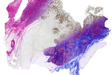
New Chemical-Free Imaging Technology Stains Tissue Samples With Light

Researchers have developed a new imaging technique that scans tissue samples with infrared light, allowing pathologists to digitally apply several different virtual stains to the same biopsy without the use of chemical dyes or stains. With this new method, researchers believe that one biopsy could yield a wide range of quick diagnostic assessments.
Traditional biopsy analysis methods require lab technicians to dip ultra-thin slices of tissue samples into a chemical bath repeatedly until pathologists can view particular cell malformations or molecules clearly. Each chemical dip risks ruining the tissue sample, and checking for more than one disease could require additional biopsy samples and dyes.
A research team led by Rohit Bhargava, University of Illinois professor of bioengineering and member of the Beckman Institute for Advanced Science and Technology, wanted to improve on this time-consuming, messy, and expensive process, according to a U. of I. News Bureau story
The new method begins in the traditional manner. Tissue samples are sliced very thin and placed under a microscope, but instead of a chemical bath, researchers used Fourier transform infrared (FTIR) spectroscopic imaging to scan the tissue and computer analysis to apply the “stain.” Once the image is scanned into the computer, a program can translate the information from the microscope into a variety of chemical stain patterns.
“Any sample can be analyzed for desired stains without material cost, time or effort, while leaving precious tissue pristine for downstream analyses,” said Bhargava.
In a study published in Technology, Bhargava’s team detailed the specifics of their technique and reproduced a number of different molecular stains by using a computer algorithm to isolate the spectra of specific molecules. Researchers explained that the computer program is a supervised learning algorithm that will expand over time using knowledge generated by chemistry.
“One of the bottlenecks in automated pathology is the extensive processing that must be applied to stained images to correct for staining artifacts and inconsistencies,” said David Mayerich, first author of the study. “The ability to apply stains uniformly across multiple samples could make these initial image processing steps significantly easier and more robust.”
The researchers demonstrated that multiple stains could even be applied simultaneously to the same image, which allowed for the creation of new images that existing methods are unable to generate.
“This approach promises to have immediate and long-term impact in changing pathology to a multiplexed molecular science — in both research and clinical practice,” Bhargava said.
Image credit: Rohit Bhargava
