New MRI Method Could Hasten Autism Diagnostics

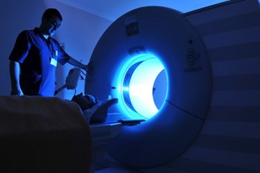
Researchers at Virginia Tech (VT) may have discovered a quicker method for diagnosing autism spectrum disorder (ASD) using magnetic resonance imaging (MRI). This new process could yield more quantitative and conclusive results than current clinical standards.
Last year, the Centers for Disease Control and Prevention (CDC) released a report that studied children in various locations across the U.S. and estimated that 1 in 68 children in those regions have been diagnosed with autism.
While some argue that this spike in numbers is due mostly to changes in how the disease is diagnosed, most scientists agree that there has been an increase in cases and that early detection is of the utmost importance, according to Time Magazine.
Current diagnostic methods for ASD rely heavily on a doctor’s judgment, the evaluation of symptomatic behaviors, and the patient’s medical history. This process is often time-consuming and frustrating for families.
The newly developed brain-imaging technique from VT researchers may help streamline the diagnostic process for ASD.
VT scientists, led by Read Montague, found that by looking for a specific marker during an MRI scan, doctors might be able to distinguish between children who have ASD and children who do not. Doctors could potentially conduct the test in under two minutes, reports the Virginia Tech News.
Montague’s research began in 2006 when his team isolated a brain function in the middle cingulate cortex that became more active when subjects were asked to take turns during an activity.
This part of the brain, researchers discovered, is responsible for a human’s ability to tell the difference between themselves and others, and it could be tracked using an MRI.
“This response is removed from our emotional input, so it makes a great quantitative marker,” Montague explains in the Virginia Tech News article. “We can use it to measure differences between people with and without ASD.”
In the current study, the VT research team showed children fifteen images of themselves and fifteen images of other children of similar age. MRI scans taken from children with ASD showed significantly less activity than children without ASD.
The VT study is not the first to use MRI scans in autism research. In December, researchers from Carnegie Mellon University (CMU) published an autism study that asked subjects to consider social activities, such as “persuade” and “hug.” The researchers then studied the subjects’ subsequent brain scans. Like the VT researchers, the CMU scientists discovered that certain brain functions could be highly accurate indicators of the disorder.
The key significance of the VT study is that the test is relatively simple can be administered quickly, which is ideal when testing small children.
In the Virginia Tech News article, Montague expressed his hopes for the broader potential for the new method: “The single-stimulus functional MRI could open the door to developing MRI-based applications for screening other cognitive disorders.”
A more comprehensive overview of Virginia Tech’s findings will be published in Clinical Psychological Science.
