Optical Fiber Provides Real-Time Anticoagulation Monitoring
By Jof Enriquez,
Follow me on Twitter @jofenriq

Using fiber optics, University of Central Florida (UCF) researchers have developed a technique to monitor, in real time, the formation of dangerous blood clots during cardiovascular procedures.
The risk of developing blood clots that can lead to a heart attack, stroke, or pulmonary embolism rises dramatically during invasive cardiovascular surgery. To lessen the risk, doctors administer anticoagulants to thin the blood, samples of which must be repeatedly withdrawn from the extracorporeal circuit (for example, during cardiopulmonary bypass) at 20-30 minute intervals throughout a procedure that can last for many hours.
Even for standard assays, such as activated clotting time (ACT) and thromboelastography (TEG), there are typical time gaps of at least 15–30 minutes, during which the exact status of blood coagulability is unknown to the surgical team.
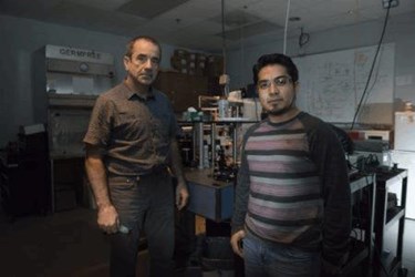
The UCF team, led by Aristide Dogariu, a Pegasus Professor in UCF’s College of Optics & Photonics, sought to eliminate these risky time gaps.
They developed an optical fiber that beams light at the blood flowing through the tubes of a heart-lung machine used in a bypass procedure. The fiber detects the light as it bounces back, through a technique called coherence-gated light scattering, and the machine detects the backscattered light to determine if red blood cells are vibrating slower than expected – a sign of increased coagulation – indicating that additional doses of a blood thinner are needed.
“It provides continuous feedback for the surgeon to make a decision on medication,” said Dogariu in a news release. “That is what’s new. Continuous, real-time monitoring is not available today."
"The method does not need sample collection, special preparation procedures or external triggering of the coagulation cascade, thus eliminating the delays associated with conventional technologies based on end-point measurements," Dogariu and colleagues added in the proof-of-concept study, published in Nature Biomedical Engineering.
They also claim that the optical fiber can be directly incorporated into existing, standard vascular access devices used in operating rooms today.
UCF's optical fiber blood monitoring technique has been tested during cardiac surgeries on 10 infants at Arnold Palmer Hospital for Children, with promising results, according to New Atlas. The team now is working to expand its proof-of-concept to a larger study.
“I absolutely see the technique having potential in the intensive care setting, where it can be part of saving the lives of critically ill patients with all kinds of other disorders,” said Dr. William DeCampli, chief of pediatric cardiac surgery at Arnold Palmer Hospital for Children, and a professor at the UCF College of Medicine.
