
Part II Of II: Applications For Hyperpolarized Gas MRI

Written by John Swartz, Duke University
Hyperpolarized gas magnetic resonance imaging (HPG-MRI) is a new technique for imaging gas-filled samples such as human lungs and porous materials. Researchers at Princeton University (Princeton, NJ), the University of California (Berkeley), and other labs are now developing the HPG-MRI technique. Recently, Nycomed-Amersham (NA; Princeton, NJ) acquired Magnetic Imaging Technologies Inc. (MITI; Durham, NC; 919-572-0954), to encourage MITI's front-line HPG-MRI work.
Medical Applications
Materials Applications
NA Acquisition Of MITI
Part I And Contacts
About The Author
Medical Applications (Back to Top)
Currently, medical imaging applications are the most popular use of HPG-MRI. Much work has been done to develop effective ways of imaging the lungs, airways, and sinuses in both animals and people. Both static imaging and dynamic flow analyses have been done using HPG-MRI.
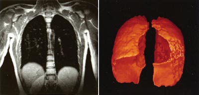
HPG-MRI can produce several different types of images, including proton images (left) and surface-rendered 3-D images. These are both from a normal subject.
Because MRI signals are sensitive to the local environment of the excited gas or water molecules, small frequency shifts in the MRI signal can be used to identify nearby materials. This phenomenon is the basis behind the well-known technique of using water-based MRI to identify fats, tissues, and bones.
Recent research has demonstrated that hyperpolarized gas MRI can be used in a similar manner. G. Allan Johnson and his colleagues at the Duke University Medical Center (Durham, NC) as well as James Brookeman at the University of Virginia (Charlottesville) have shown that helium-3 MRI images of lungs can differentiate healthy and diseased lung tissue. Because of this, HPG-MRI could be a useful tool for diagnosing emphysema, chronic obstructive pulmonary disorder, and other lung diseases. Researchers are also studying the efficacy of using HPG-MRI to image the colon and detect colon cancer. Currently, MITI and NA are waiting for approval from the U.S. Food and Drug Administration (FDA) to conduct HPG-MRI medical trials on a wider basis.
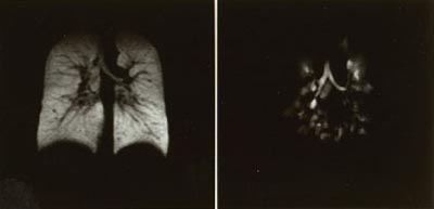
These 3He HPG-MRI images clearly show the difference between healthy and diseased lung tissue. The left image shows a normal subject, while the right image shows a subject with chronic obstructive pulmonary disorder.
Materials Applications (Back to Top)
Hyperpolarized gas MRI has also been used in non-destructive materials evaluations. Researchers at Princeton University have demonstrated that hyperpolarized MRI signals are different at the edges of a container than at the center. Additional research is now underway.
At the University of California (Berkeley), Alex Pines and his colleagues are using the MRI of hyperpolarized xenon-129 to study gas flow through porous materials, called aerogels. These materials have sub-micron channels and voids. Because hyperpolarized MRI studies provide a new means of studying gas transport and flow characteristics in materials, they are ideal for finding new applications for aerogels. Although preliminary, these analysis techniques show that hyperpolarized gas MRI could be a useful tool for studying porous and hollow structures, as well as fibrous materials.
At the National Institute of Standards and Technology (NIST; Gaithersburg, MD) researchers are using hyperpolarized helium-3 to measure and produce polarized neutron beams that can analyze materials. Neutron-beam work at NIST includes studying heavy metals in lake samples, characterizing new biomimetic materials, and studying conformational changes in proteins. The HPG-MRI technology is likely to impact these research areas if neutron beams are enhanced with hyperpolarized helium.
Another advantage of HPG-MRI is that unlike conventional MRI, hyperpolarized gases remain polarized without a strong magnetic alignment field. This means that it is possible to use hyperpolarized gases to make MRI images without the large static magnets that are required for traditional MRI. Ronald Walsworth and his colleagues at the Harvard-Smithsonian Center for Astrophysics (Cambridge, MA) have recently built a MRI system that uses small magnets. Walsworth hopes to use the system for practical low-field imaging. In addition, Walsworth's group has studied the dynamics and physics of hyperpolarized liquid xenon. Potential applications include studying convective flow in fluids and studying interfacial shears in fluids.
NA Acquisition Of MITI (Back to Top)
On Aug. 5, 1999, Nycomed-Amersham (Princeton, NJ), a leading provider of radiotherapy products and in-vivo diagnostic imaging, acquired Magnetic Imaging Technologies Inc. (MITI; Durham, NC). For the past five years, MITI has worked to develop the HPG-MRI technology.
Although primarily aimed at medical imaging and diagnostic applications, HPG-MRI may be a new tool for researchers interested in techniques in non-destructive analysis and testing. For the immediate future, NA-MITI will continue to focus on bringing HPG-MRI to the medical market, conducting clinical trials, pursuing regulatory approval, and developing the technology for clinicians.
Because NA has built a sophisticated distribution network for short-lived (few hours to a few days) radioisotopes, it is likely that NA will facilitate HPG-MRI research by distributing hyperpolarized gases on request. This will probably be a more cost-effective approach than selling individual researchers and clinicians the hardware and support needed to produce hyperpolarized gases.
Currently, HPG-MRI is an infant technology for which new applications and techniques are still being discovered. The NA acquisition of MITI should provide some stability to the young commercial venture into HPG production and hardware, eventually making HPG-MRI accessible to a wider group of users.
Part I And Contacts (Back to Top)
For more information about how MRI and HPG-MRI work, please read Part I Of II: Hyperpolarized MRI Images Gas-Filled Samples.
For more information, contact Bastiaan Driehuys at MITI (now Nycomed-Amersham Imaging); 2500 Meridian Parkway, Suite 175; Durham, NC 27713; Tel: 919-572-0954 ext. 222; Fax: 919-572-0689; e-mail: driehuys@miti2.com.
Carol Perlman at Nycomed-Amersham Imaging; 101 Carnegie Center; Princeton, NJ 08540; Tel: 609-514-6000; e-mail cape@americas.nycomed.com
James Brookeman, professor of radiology and biomedical engineering, University of Virginia; Charlottesville, VA 22908.
Ronald Walsworth at the Harvard-Smithsonian Center for Astrophysics; MS 59; 60 Garden St.; Cambridge, MA 02138.
About The Author (Back to Top)
John Swartz received his Ph.D. in electrical engineering from Duke University in 1994. Currently he is a research associate in physics and biomedical engineering at Duke University studying laser physics, medical applications of lasers, and applied optics. Dr. Swartz is also an adjunct researcher in the Physics Department at Dartmouth College, investigating far infrared physics and free-electron laser applications.