
RSNA 1999: Diagnostic Imaging Equipment Manufacturers Unspool Digital Capabilities

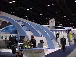
Nearly 60,000 physicians, radiology professionals and diagnostic imaging equipment manufacturer representatives converged at Chicago's McCormick Place last month for the 1999 Scientific Assembly and Annual Meeting of the Radiological Society of North America.
Armed with their mental shopping lists, attendees were able to scout a wide variety of diagnostic imaging modalities and related radiology products from 630 different companies occupying more than 400,000 square feet of commercial exhibit space.
Although anything digital seemed to captivate showgoers, doctors and other radiology professionals were able to inspect a litany of other products, too. They include traditional and open magnetic resonance imaging units, computed tomography scanners, mammography systems, nuclear medicine units, ultrasound units, computed radiography devices, as well as megabytes of software offerings and stacks of published material.
In the first of two reports, Hospital Network.com provides highlights of selected products making waves at the show. In the second report, we'll show you what some of the largest radiology players in the market displayed.
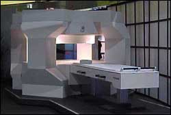
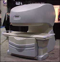
One of the most avid promoters of open magnetic resonance imaging technology, FONAR Corp. (Melville, NY) showcased its seven MRI scanner products, two of which made their debut at the show, and displayed high-resolution images produced from each as well.
The Echo and The Pinnacle were the two newest additions to FONAR's expanding line. The small, compact Echo with its 0.35 Tesla magnet is being aimed at facilities with high-end MRI scanners but are looking for a low-cost model to complement their operations. It carries a $630,000 list price tag. The larger and faster Pinnacle, which has been under development and is scheduled for market release by fourth quarter 2000, contains a 0.8 Tesla magnet. Daniel Culver, FONAR's director of communications, called The Pinnacle "the only high-field superconducting open MRI scanner on the market."
FONAR also demonstrated three of its more creative open MRI units: The Stand-Up MRI, the OR 360° MRI and the Open Sky MRI.
From its Stand-Up MRI with a 0.6 Tesla magnet, FONAR generated movies (dynamic motion cines) of the lumbar spine, the cervical spine and joints while a patient stood upright in the weight-bearing position. The company said patients could be scanned while standing, sitting, reclining at any angle or even bending to touch their toes. The 0.6 Tesla OR 360° MRI is a dual-purpose unit made for MRI-guided surgical and interventional procedures in the operating room as well as for conventional diagnostic scanning. Sans a four-post iron frame, it gives clinicians total access to the patient. The Open Sky MRI is designed more for patient comfort. It's simply the OR 360° MRI in a room painted with a panoramic mural.
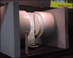
FONAR also exhibited "enhanced" versions of its QUAD 12000 and QUAD 7000 open MRI scanners that are its signature units for generating high-speed, high-resolution, high-contrast scans. The QUAD 12000 contains a 0.6 Tesla magnet; the smaller QUAD 7000 uses a 0.35 Tesla magnet.
PhorMax Corp. (Palo Alto, CA) unveiled its CRView desktop radiography scanning system, which works with a facility's existing X-ray equipment and procedures. CRView consists of a desktop scanner and a Windows-NT personal computer workstation that runs a proprietary software application for the acquisition, manipulation and communication of digital radiographs. It captures images on a reusable phosphor-based imaging plate that's housed in a cassette about the same size as an X-ray film cassette. After capturing the image, CRView automatically converts it to a digital format and then erases the plate for reuse.
PhorMax plans to make CRView available for purchase in mid-year 2000 for about $50,000, compared to the $75,000 to $200,000 for larger computed radiography units.
Miriam Bischoff, senior product manager, told Hospital Network.com that CRView is good for facilities that aren't ready to go fully digital but don't want to rely solely on film, occupying a middle ground between the two. Because the product makes direct filmless 2-D radiographs right at the point of care, PhorMax is eager to promote it for hospital emergency and operating rooms, Bischoff said.
"[CRView] is a neat little application that addresses the low-volume facilities who can't justify purchasing a full-fledged CR system," said Niklaus Fincher, vice president, business development, InPhact Inc. (Brentwood, TN), a technology management services company specializing in diagnostic imaging. "You can scan and clean the plate cassette in fewer steps and it's automated."
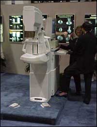
Fischer Imaging Corp. (Denver) displayed its line of mammography systems, including its new full-field digital mammography unit that awaits Food and Drug Administration marketing clearance.
Fischer Imaging's SenoScan full-field digital mammography system is an outgrowth of its Mammovision digital imaging system, which uses a single fiber optic taper bonded to a charge-coupled device (CCD) chip for stereotactic breast biopsies. SenoScan employs a larger field-of-view imaging platform that's designed to improve image quality, reduce patient radiation exposure and enhance patient comfort and positioning.
Fischer Imaging also showed its flagship Mammotest Plus and the newer Mammotest Plus/S prone stereotactic breast biopsy systems. Although both use an interchangeable portable digital camera and provide direct needle access to a lesion in any area of the breast, the newer model's needle is maneuvered electronically using a touch-pad control box. It also offers remote targeting capabilities and will enable add-on integrated ultrasound as a future upgrade.
Ethicon Endo-Surgery Inc. (Cincinnati) demonstrated its new Mammotome system for minimally invasive breast biopsies. Ethicon Endo-Surgery's older breast biopsy technology, which the Johnson & Johnson company credited as the first hand-held breast biopsy system, involved a larger gauge needle that was manually inserted by the doctor while the patient remained under local anesthesia. However, the smaller gauge needle is software-driven in the Mammotome system, which includes a computer-controlled vacuum system with a high-speed rotating cutter. Doctors can insert the needle using a hand-held computer and only have to do it once, unlike the traditional spring-loaded biopsy systems that require multiple insertions. The system also allows the doctor to deploy a tissue marker in the biopsy cavity for any follow-up care.
Tyco Healthcare Group's U.S. Surgical division (Norwalk, CT) promoted its own vacuum-assisted breast biopsy system, MIBB (Minimally Invasive Breast Biopsy) and the upcoming faster MIBB 2. Both systems use computer-controlled stereotactic guidance and boast of drawing larger specimens than the competition's products. MIBB also employs a site marker device to leave clips in the biopsy cavity.
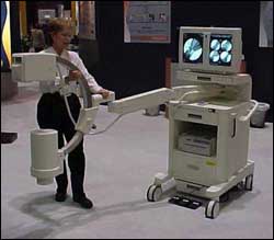
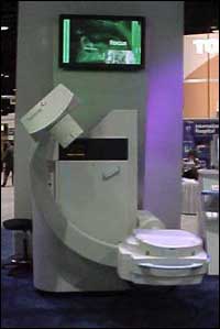
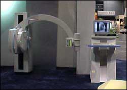
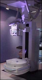
Hologic Inc. (Bedford, MA) and its two subsidiaries Direct Radiography Corp. (Newark, DE) and FluoroScan (Northbrook, IL) showcased its broad array of direct-to-digital radiographic and fluorscopic systems and bone densitometers.
Hologic's new Delphi QDR series bone densitometers look like they were just beamed off the Starship Enterprise. The Delphi units (QDR 4500 and QDR 4500 Elite) enable doctors to assess two of the strongest risk factors for osteoporotic fractures – existing vertebral fractures and low bone density in the hip, spine and forearm areas. The point-of-care system uses a 10-second, low-dose single-energy X-ray image for doctors to evaluate any vertebral deformities. Armed with its auto analysis capabilities using Windows software, the high-resolution detector arrays can scan and evaluate both hips in a single exam. Several of the QDR models can perform whole body scans. Using amorphous selenium rather than cesium iodide, they can produce digital radiographic images in seconds so doctors can bypass X-ray film and chemicals.
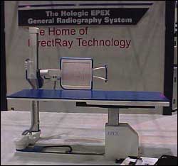
Direct Radiography, or DirectRay for short, displayed its new top-of-the-line Hologic EPEX direct-to-digital general radiography system and its more basic Hologic RADEX model. Both systems are scheduled for rollout in second quarter 2000. DirectRay is targeting the low-end Hologic RADEX for the outpatient surgery market. Hologic EPEX will carry a $395,000 list price tag; Hologic RADEX will sport a less-than $330,000 list price. These two systems join DirectRay's DR1000C digital chest system and its DR1000 general radiography system for trauma, both of which were introduced two years ago.
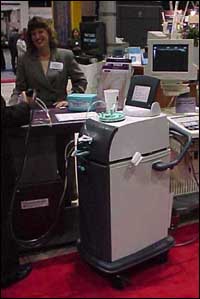
Fluoroscan representatives demonstrated the maneuverability and user-friendliness of the company's Premier mini-C arm, which originally debuted in 1998 but was upgraded last year. The upgrade includes a remote-control keypad that instructs the user on how to operate the unit. It also includes an audit log that tracks usage by physician, patient and procedure.
Premier's iso-centered C-arm rotates 360°, and its SureLock feature locks the C-arm securely into any position around the surgical patient. The flex arm holding the C-arm in place can extend 47 inches from the mobile computer cart, which also carries dual monitors (the right one allows up to four images to update in turn), keyboard, disk storage and retrieval unit and printer.
With its Jetsons-inspired architectural design, the futuristic "Competence Center" from Swissray Medical AG (Hochdorf, Switzerland) housed the company's ddR Systems, including the ddR Multi-System, ddR Chest System and the new ddR Combi direct digital radiography units. It also featured rows of flat-panel display monitors for doctors to view diagnostic imaging results from Swissray products.
The ddR Multi-System performs all direct digital radiographic exams on recumbent, sitting and upright patients, eliminating the need for a separate skull or chest imaging system. Doctors operate the unit either by remote or by push buttons on the unit itself.
Swissray's ddR Chest-System, dubbed the "workhorse" because chest radiography typically accounts for more than 50% of a department's workload, allows a full-sized chest image to be acquired every five seconds. The system also provides immediate digital diagnostic images for review on a computer workstation.
The new ddR Combi has a motorized overhead tube crane that can be operated manually as well. It can be moved to allow unrestricted patient access through remote-controlled, dual-speed motor-driven positioning. In fact, the ddR Combi Solo provides a complete digital imaging chain – generator, tube, collimator, digital detector, image review workstation and algorithms – that can be retrofitted to an existing tube crane system or floor-mounted tube stand.
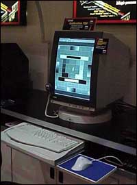
DOME Imaging Systems Inc. (Waltham, MA) introduced the Calibration TQA (Total Quality Assurance) for Windows 95 and NT 4.0 operating systems. DOME's new product, which focuses on enterprise-wide applications supporting multiple monitors, evolved from its three-year-old Luminance Calibration System, which focused on individual workstations. Cal TQA includes a new photometer that requires the user to hold the sensor to the monitor screen and software. Also users can operate Cal TQA either in standard or service mode. In standard mode, users can perform easy calibration and conformance testing through clearly defined, step-by-step procedures. In service mode, users can monitor the performance of workstations, setting tolerance levels and configuring the system to notify users with service alert and failure warnings when luminance values fail to meet specified tolerances.
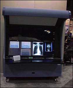
Two years ago, RSNA attendees discovered a digital alternative to the conventional light box with which view X-ray film images. SmartLight Inc. (Hackensack, NJ) introduced its Digital Film Viewer to an audience captivated by the device's image quality and its ability to display smaller, lower contrast radiographic objects. At the 1998 RSNA conference, SmartLight presented study results from more than 1,000 radiologists from 18 countries who found they could detect lesions 25% to 50% smaller in volume and fainter using SmartLight's device than what they could using traditional light boxes. At the 1999 RSNA conference, SmartLight rolled out the newest enhancement to its product – automation.
Radiology professionals were able to try out SmartLight's new entry-level SL8000 SLX Smart Motorized Viewer. The SLX features a fast-moving films belt controlled by keypad and foot pedals and provides fully automatic masking with adaptive illumination and scatter suppression. Radiologists also can configure the SLX to be a computerized film archive manager for study-by-study film viewing and referral.
Millennium Technology Inc. (Richmond, British Columbia) demonstrated its Virgo MRI, ergonomically designed for whole-body imaging. A mid-range open MRI with a permanent 0.35 Tesla magnet in a "C" shape, Virgo also includes a fully motorized detachable table for more efficient patient maneuvering.
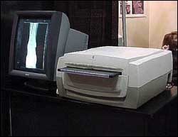
VIDAR Systems Corp. (Herndon, VA) introduced its new Sierra film digitizer, which the company called the first model priced under $10,000. VIDAR's Sierra weighs only 15 pounds and can be wall-mounted because of its size; it's slightly larger than a one-panel light box. Typically, film digitizers weigh between 50 pounds and 200 pounds. The Sierra also includes a continuous calibration feature that enables the unit to make high-level grayscale reproductions and enhances image quality and it includes a six-sheet film feeder to automate the process.
Voxar Ltd. (San Antonio, TX) showed its computer-based three-dimensional medical visualization software. Called Plug ‘n View 3D, the software complies with the Windows operating system as well as the Digital Imaging Communications in Medicine (DICOM) standard. The company aims to target its $5,000 3-D software package to compete against Unix-based 3-D workstations.
Surgi-Vision Inc. (Atlanta) demonstrated how the company's Endo-Esophageal Magnetic Resonance Imaging (EEMRI) coil provides physicians clear images of the heart during radio-frequency ablation, a procedure used to treat rapid, uncoordinated heartbeats. To treat atrial fibrillation, doctors insert a catheter into the heart through a vein in the leg and then burn heart tissue with radio-frequency energy. But applying energy to the heart's inner surface must be precise (within an area that is 1/5th of an inch) to be effective. Using an internal MRI coil, Voxar researchers found they could easily distinguish the ablated areas of the heart from the unaffected areas.
Algotech Systems Ltd. (Raanana, Israel) unveiled MediPrime, its new integrated primary radiology reading and reporting workstation and introduced a number of upgrades and enhancements to its ImagiNet products. MediPrime builds sophisticated worklists and automatically calls up studies to be read on high-resolution monitors. It also searches for and displays all previous patient studies and relevant clinical data on all DICOM devices in a facility's system. Algotech's MediFlow workflow management tool now includes D-Route (DICOM-Route) smart agents, which automatically send important patient files where needed throughout the system.
MediSurf, Algotech's Web-based access engine, now includes a "data push" feature to distribute clinical information online to appropriate medical professionals. Also new to MediSurf is SurfLink, which is an online conferencing tool that allows two users to consult about a study. Finally, Algotech debuted version 4.0 of its ProVision multi-modality processing station, which offers Web-based image accessing capabilities and 3-D internal anatomical renderings from mathematically complex volumetric image data. ProVision also added volume rendering-based virtual endoscopic and dedicated virtual colonoscopic tools that use computed tomography and MRI studies to explore body cavities.
Canon U.S.A. Inc. (Lake Success, NY) introduced its new Canon DR Bucky Sensor Unit System CXDI-22 digital radiography (DR) system, which provides clinicians with immediate X-ray preview images that can be transmitted from an operation panel for viewing, printing or archiving. The CXDI-22 features a 17-inch-by 17-inch imaging area, enabling images to be displayed in portrait or landscape orientation. Carrying a $200,000 list price, the CXDI-22 is scheduled to be available in January 2000.
Medweb (San Francisco) demonstrated its MedwebSAT distributed telemedicine product. MedwebSAT is a low-cost, high-bandwidth satellite service that links multiple facilities in North America, Mexico, Hawaii and the Virgin Islands, enabling on-call radiologists and referring physicians to transmit patient studies and other medical records. Medweb offers similar systems to cover facilities in Southeast Asia and Europe. MedwebDSL allows clinicians to connect from their homes.
ADAC Laboratories (Milpitas, CA) introduced the Skylight system, a gantry-free nuclear medicine gamma camera. Skylight, which isn't yet ready for market, allows gamma detectors to be mounted directly into a room structure. The detectors can be moved in any direction -- either together or independently -- to accommodate the patient position rather than having the patient positioned to fit the camera. ADAC also is looking to expand Skylight's use so that it can be combined it with other diagnostic imaging modalities such as CT or ultrasound or used in surgical and interventional applications.
For more information on these and other radiology products, visit Hospital Network.com's continually updated Product Showcase.