
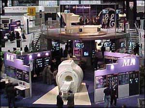
RSNA 1999: Radiology Equipment Giants Keep Clinicians Busy with Many Demos

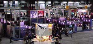
Doctors and other radiology professionals swarmed around the islet-sized booths of the larger diagnostic imaging equipment manufacturers in an effort to glimpse what the market's elite players were promoting during the 1999 Scientific Assembly and Annual Meeting of the Radiological Society of North America at Chicago's McCormick Place last month.
With the vast array of diagnostic imaging modalities and related radiology products on display, attendees were not disappointed. In the final report from RSNA 1999, Hospital Network.com offers product highlights from some of the market leaders who occupied considerable real estate in one of the two exhibit halls.
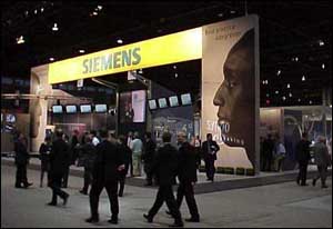
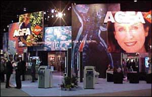
As attendees streamed into McCormick Place's North Building, they came face-to-face with the twin towers of that exhibit hall – the booths of Agfa Corp. (Ridgefield Park, NJ) on the right and Siemens Medical Systems Inc. (Iselin, NJ) on the left.
Agfa showcased its Picture Archiving and Communications Systems (PACS) capabilities, particularly through its IMPAX line. In an effort to introduce the IMPAX enterprise PACS system to the mainstream market, Agfa displayed its IMPAX Basix, an entry-level version of its new IMPAX R4 system. IMPAX Basix is designed to meet the needs of smaller or lower-volume healthcare facilities, such as 100-bed hospitals. Although scaled down to support fewer modalities and displays, IMPAX Basix is highly modular. It can support multiple software modules on an appropriately scaled Windows NT hardware platform and can be upgraded to a full IMPAX system.
IMPAX R4 is the fourth version of the enterprise PACS software, which now features a faster and more flexible user interface complete with custom worklists, as well as a more flexible architecture and more powerful image compression capabilities. New to IMPAX is a voice-recognition and digital dictation software.
Agfa announced it co-launched a new company, called IMPAX Technology Inc., with Mitra Imaging Inc. to develop software applications for the IMPAX line. The first of two products to emerge from IMPAX will be the new Embedded-IS option that will turn IMPAX into an integrated imaging and information system for the radiology department. It's slated for launch in mid-2000. The second product involves extending the capabilities of IMPAX to hospital cardiology departments. The rollout is slated for second quarter 2000.
Agfa also showed its new COMPACT E.O.S., combining the company's daylight film handling system with a dedicated Classic E.O.S. processor. The unit reduces power consumption, noise and heat as well as wastewater silver content below 1 part-per-million and it enables users to consume 35% less fixer.
Other products Agfa introduced were its Agfa Diagnostic Center (ADC) automated digitizer, which converts X-ray film images into a digital format, its new HD Mammography system with high-definition screens, and its Drystar 4000 series of direct thermal, black and white compact floor standing printers. The first model, Drystar 4500 is targeted for availability in mid-2000 with the Drystar 4500M and 4700M slated for release at the end of 2001.
Across the aisle, Siemens led radiology professionals on a tour of a virtual hospital equipped with many of its new products. Siemens' theme: Using information technology to save time and money without compromising the quality of patient care. In the Hospital Administration/Patient Registration area, attendees viewed an enterprise-wide clinical information system and electronic patient records at work, along with a radiology information system linked with the company's Sienet PACS.
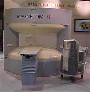
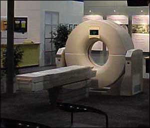
In the magnetic resonance imaging area, Siemens displayed its Magnetom Open viva MRI unit, which is designed for high-field applications, patient comfort and fast imaging results. Although it's not yet commercially available, the Magnetom Open viva includes permanent magnet technology in a C-shaped design. The company also introduced a software platform for its MRI products called Mrease. The software combines Siemens "syngo" software architecture for all imaging modalities with its trademarked Integrated Panoramic Array, which has been expanded to enable remote patient handling.
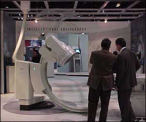
In the angiography area, Siemens showed its Angiostar unit, which is a combined system for angiography and cardiography. The company's new 3D Virtuoso workstation can reconstruct Digital Subtraction Angiography images in three dimensions, allowing physicians to view vascular structures in depth from any angle. Although 3D Virtuoso was originally designed to provide instant access to computed tomography and MRI data in 3-D, the workstation also can automatically transform conventional X-ray images from Siemens' Multistar Plus, Angiostar Plus and Neurostar Plus systems into 3-D images in roughly five minutes.
In computed tomography, Siemens displayed its new mid-priced Somatom Balance and Emotion CT scanners, along with its Volume Zoom multi-slice capability upgrade. In ultrasound, Siemens introduced its new mobile Sonoline Adara as well as its flagship Elegra and Omnia and Prima models. The company also touted its Siemens Photopic Ultrasound Imaging technology, which recognizes the acoustic "fingerprint" of a standard, grayscale ultrasound image, balances it and then translates it into a color-enriched image that can be viewed in bright-light or photopic conditions.
In the nuclear medicine area, Siemens demonstrated its new PC-based workstation called e.station 2000 to be used with its E.CAM group of gamma cameras. Through the Windows NT operating system, physicians using e.station 2000 can integrated data acquisition, reconstruct images and configure displays however they choose. Data also can be distributed automatically to a wide variety of DICOM-based systems, including PACS, archives, printers and other imaging workstations.
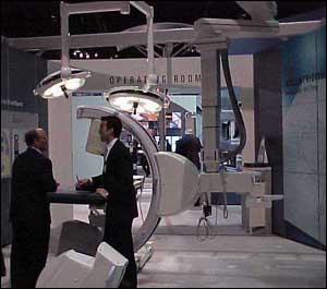
Siemens emphasized how imaging can be used in the operating room during surgery. The company displayed its ceiling-mounted Arcoskop C-arm as well as its Siremobil Iso-C model. As part of its network-connected virtual hospital, Siemens also designated areas in radiography, fluoroscopy (showcasing its Iconos R200 and Fluorospot systems), breast imaging (showcasing its Mammomat line), oncology, ward and reading and service rooms.
Walking deeper into the North Building and down the aisle from Agfa, attendees viewed products being exhibited by Philips Medical Systems (Shelton, CT) and its ATL Ultrasound (Bothell, WA) unit in a smaller adjacent booth.
Making a big splash in the Philips booth was its new Gyroscan Intera MRI, which the company noted lets physicians replace traditional exams with faster, less costly and more informative MR studies. Gyroscan Intera sports a short gantry and is equipped to handle a broad range of exams, including neurological, musculoskeletal and abdominal imaging, functional and cardiac studies, contrast-enhanced angiography and spectroscopic imaging. In addition to the general-purpose Gyroscan Intera system, Philips also unveiled the Gyroscan Intera CV, which is designed for cardiovascular applications, and the Gyroscan Intera I/T, which will be configured for interventional and therapeutic applications and awaits Food and Drug Administration marketing clearance.
The Gyroscan Intera boasts a number of applications. They include:
- A high-resolution contrast-enhanced peripheral angiogram that can be done in a couple of minutes on an outpatient basis
- A comprehensive cardiac evaluation in a single one-hour MR session on an outpatient basis
- Works-in-progress interventional and therapeutic procedures, including biopsies, ablations, targeted drug delivery therapy and tumor resections
- A comprehensive stroke examination completed in less than six minutes covering all essential scan sequences
- A total body diagnostic capability.
Philips also promoted its new CT Vision systems – the CT Aura and the CT Secura – both of which enable doctors to perform a full range of studies from the routine to the complex. The Aura and Secura feature full rotation sub-second helical scanning and reconstruction times, advanced post-processing and dose-management software and a dual-monitor, dual-keyboard operator console.
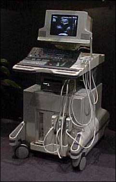
Next door to Philips, ATL Ultrasound showcased its new HDI 3500 system, a mid-range ultrasound unit between its low-end HDI 3000 system and its top-of-the-line HDI 5000 system. The HDI 3500 is based on the platform of the low-end system but it incorporates the advanced processing capabilities and user interface technologies of its premium system. The HDI 3500 features 256 digital channels and high-performance signal processing technology. It also has a new user-centric control panel and sits on four-wheel swivel castors, as does the HDI 5000.
ATL also featured its new SonoCT Real-Time Compound Imaging ultrasound technology, which the company promoted as being able to compile up to nine times the information of conventional ultrasound.
Compound imaging, which is common in CT and MRI, obtains images from different viewing angles, then combines them into a single image. But conventional ultrasound machines didn't have the computer power to do this in real time. ATL noted that this new technology can be used to identify smaller lesions sooner, particularly in the breast area. The company began developing the technology to eliminate image interference, such as speckle and refractive shadows, which make seeing the image more difficult and also affects a doctor's diagnosis.
ATL is making the SonoCT Real-Time Compound Imaging technology available on its HDI 5000 system.
Over in McCormick Place's South Building, GE Medical Systems (Milwaukee) showcased its latest offerings in MRI, CT, nuclear medicine and positron emission tomography (PET), ultrasound, mammography, X-ray, PACS and imaging software as part of its "Destination Digital" theme that stretched across its entire multi-modality product line.
Among the MRI units GE Medical promoted within the MRI tower at one end of its booth was the new GE Signa OpenSpeed high-field open MRI system, which the company said can scan patients three times faster than competitive systems. GE Medical said the development of the new open MRI cost more than $30 million. The company expects to install up to 100 of them around the world by the end of 2000.
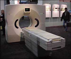
GE Medical also promoted the results of its year-old LightSpeed QX/I multi-slice CT scanner, of which 200 units have been installed throughout the world. By scanning multiple adjacent slices of the anatomy in less than one second, the LightSpeed CT captures multiple images six times faster than traditional single-slice CT scanners.
The LightSpeed CT also gives physicians the ability to use thinner slices for every exam to obtain more detailed diagnostic information. Because physicians like to view every scan in 3-D but can't due to time constraints, LightSpeed's Direct 3D capability solves the problem by rendering three-dimensional volume images concurrently with the two-dimensional axial images. With the SmartTools software, radiologists can automate every step of the exam.
GE Medical added two new products to its CT HiSpeed line – the HiSpeed ZX/I and the mobile version HiSpeed DX/i M. With more advanced helical coverage and an improved X-ray tube, the HiSpeed additions are designed to generate faster scanning speeds.
In its nuclear medicine/positron emission tomography (PET) pavilion, GE Medical introduced its GE Mobile PET Advance unit. PET scanners are used to detect signs of certain diseases, such as cancer, at a much earlier stage than other imaging modalities. The unit also includes a wireless data transceiver that can electronically share patient data without the need for traditional network connections.
At the opposite end of the MRI tower, GE Medical displayed its ultrasound units, giving specific weight to its new GE Logiq 500 Pro "smart" ultrasound system. The GE Logiq 500 Pro enables radiologists to display high-quality anatomical images at the touch of a single button.
In the company's Women's Healthcare section adjacent to the ultrasound display, GE Medical promoted its GE Senographe DMR+ mammography system. It features a specially designed GE X-ray tube that produces more accurate images during the initial exam but with 40% less radiation. That's because it features a special viewing option that lets doctors take preliminary images before pressing the exposure button.
GE Medical also noted that the new mammography system complies with all equipment requirements of the FDA's Mammography Quality Standard Act (MQSA).
Next door in the digital X-ray/X-ray pavilion, GE Medical showed its GE Advantix LCA+ system, which is designed to help doctors diagnose blood vessel problems (such as peripheral vascular disease) despite reducing contrast injections and X-ray exposure to patients.
Doctors can use the GE Advantix LCA+ to pinpoint the location of blood vessel blockages and to guide the placement of the balloon during angioplasty.
Other features of the unit include a 3-D imaging capability and the ability to take pictures of both legs simultaneously.
GE Medical touted a number of software offerings to help integrate imaging procedures, including improvements to its PathSpeed PACS product. The GE RadStore allows hospitals to digitally store and transmit patient images and information through a centralized digital archive.
More compelling is the new GE iDrive, a fully interactive imaging software that lets doctors take self-guided, real-time tours inside patients – even when the patient is breathing and moving.
The software takes quick, continuous images of the patient's anatomy after he or she enters the MRI magnet. Once the doctor detects a lesion, the software provides even more detailed information about the location within the organ so the doctor can study the progression of diseases, recommend surgery options and plan treatment programs. The software also measures how long it takes for a contrast agent to move from the point of injection to the blood vessel being studied.
Toshiba America Medical Systems Inc. (Tustin, CA) displayed a wide range of product modalities, spanning MRI, CT, ultrasound and nuclear medicine.
Toshiba unveiled its Excelart 1.5 Tesla MRI, touting it as the "quietest and widest MRI system in the world." Excelart features a short-bore design and a 655-millimeter opening – wide for a closed MRI system. The unit's Pianissimo software reduces examination noise by up to 90%.
But Toshiba also highlighted its Opart open MRI system, promoting several enhancements to the product. Those enhancements include a fully motorized multi-directional table, four new multi-channel R/F coils, an in-room fluoroscopy monitor, new operating software and DICOM management capabilities and high-field applications.
In the CT area, Toshiba announced its Aquillon and Asteion multi-slice CT scanners. Aquillon is designed to deliver one-half second, full rotation, multi-slice images, or eight slices per second. Asteion performs 0.75-second full-rotation scans.
Toshiba also promoted its all-digital PowerVision 8000 ultrasound system, which is geared for usage in multiple hospital departments. The system has 512 channels configured for whole-body, radiology and cardiology imaging applications. It supports all DICOM service classes and integrated 3-D and Stress Echo applications.
Toshiba demonstrated its new picture archiving communication system called simPACS, which runs on a Windows NT-based platform.