Scientists Develop Imaging Software To More Accurately Diagnose Lung Cancer
By Joel Lindsey

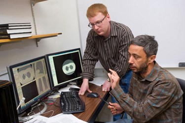
A research team led by scientists at the Rochester Institute of Technology (RIT) in New York is developing medical imaging software to measure the growth of nodules in patients at risk for lung cancer.
The project is aimed at overcoming some of the most significant challenges of analyzing and interpreting computed tomography (CT) scans, enabling much more accurate and earlier cancer diagnoses, as well as decreasing the need for biopsies.
According to an RIT press release, there are a number of persistent problems that complicate the use of CT scans as a way of tracking nodule growth. Discrepancies between images generated from different scans can arise from changes in a patient’s physical positioning, including whether or not the patient was inhaling at the time the image was created. Similarly, weight gain between scans can significantly alter the appearance of nodules in a CT scan. Such disparities between images can make it challenging for radiologists to accurately interpret what they see.
“Having even 1 or 2 millimeters of difference could throw off the estimates of the volumes of the nodules because the size of the nodules might be 5 millimeters or so,” said Nathan Cahill, an associate professor at RIT’s School of Mathematical Sciences who was involved with the research. “The goal of this project is to develop an algorithm that tries to compensate for all those potential background factors.”
The research team is developing a computer program that will align image backgrounds of CT scans in order to create a common frame of reference between sets of images. In the end, this could make it possible to more accurately assess whether changes between images indicate dangerous nodule growth or are simply background noise.
“Then we can estimate the volumes, which will allow us to more accurately estimate the doubling time and have a better chance to determine if it’s a malignant growth or benign,” said Cahill.
The next step for the technology is clinically validating the algorithm’s results. The team will review 30 CT scans of patients who were treated for lung cancer at the University of Pittsburgh Medical Center.
“With today’s technology we have the ability to create three-dimensional datasets, volumes of image data that can be manipulated and analyzed in non-visual ways,” David Fetzer, a radiologist at the University of Pittsburgh Medical Center who is collaborating on the project, said. “With techniques such as this we may be able to compensate for background changes and, hopefully, more accurately show growth, assess aggressiveness, or prove stability of a nodule. This accurate assessment could dramatically affect patient care, decrease cost and the number of unnecessary procedures, and improve outcomes through earlier cancer detection.”
Image credit: A. Sue Weisler / Rochester Institute of Technology
