Water-Repellent Nanosheet Wrapper Enhances Biological Microscopy
By Jof Enriquez,
Follow me on Twitter @jofenriq

Japanese scientists have developed a unique water-repellent nanomaterial that can be wrapped around biological tissue to visualize high-quality images for a longer period of time.
Inspired by plastic food wraps, researcher Yosuke Okamura from Tokai University and colleagues explored the effectiveness of a type of organic, highly optically transparent nanomaterial, commercially known as 'CYTOP', to fix tissue samples and generate high-resolution microscopic images.
Okamura and his team said their research "establishes and verifies the superiority of nanosheet wrapping mounts for tissue imaging," according to a news release from the Micro/Nano Technology Center, Tokai University.
The researchers were trying to solve a known limitation in biological microscopy technology where tissue samples dry out and degrade quickly, which results in poor images and inaccurate characterization of samples.
Okamura's team had known that CYTOP is exceptionally water-repellent and can easily stretch into nanosheets with surfaces free of cracks or wrinkles. Because of these properties, the researchers believed that CYTOP could function effectively as wrapping for biological material.
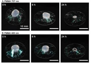
In an experiment, they coated a biomaterial sample called an alginate-hydrogel in a CYTOP nanosheet to check how it retains water. Results showed that, after 24 hours, the 133-nanometer-thick nanosheet retained as much as 60 percent of the amount of water it held at the start of testing, which gave it enough surface adhesion to fix tissue samples. Moreover, the rate of water retention rose in proportion to nanosheet thickness, when adjusted.
By contrast, another sample without the CYTOP wrapper dried out totally after only 10 hours.
The team proceeded to confirm the results using an actual biological sample: 1-mm thick brain slices from mice, exhibiting enhanced expression of yellow fluorescent protein for imaging purposes.
In this test, water evaporation caused local, non-uniform sample shrinkage of tissue samples without CYTOP wrapping, leading to a blurred image of the brain slices. Samples coated with CYTOP, on the other hand, yielded high-resolution images scanned from a large area (more than 750 µm x 750 µm) over a long time (about 2 hours).
"By wrapping cleared mouse brain slices with a 133 nm thick CYTOP nanosheet, this study achieves high spatial resolution neuron images while scanning over a large area for a long period of time. No visible artifacts arising from sample shrinkage can be detected," the scientists wrote in the 11 August 2017 issue of Advanced Materials.
Beyond two hours, however, shrinkage of tissue eventually manifest, the researchers said. To address this issue, they propose a common technique in biological microscopy which embeds the sample with agarose, a gel-forming material, providing a stability matrix.
